Penicillium
Please be patient while images download

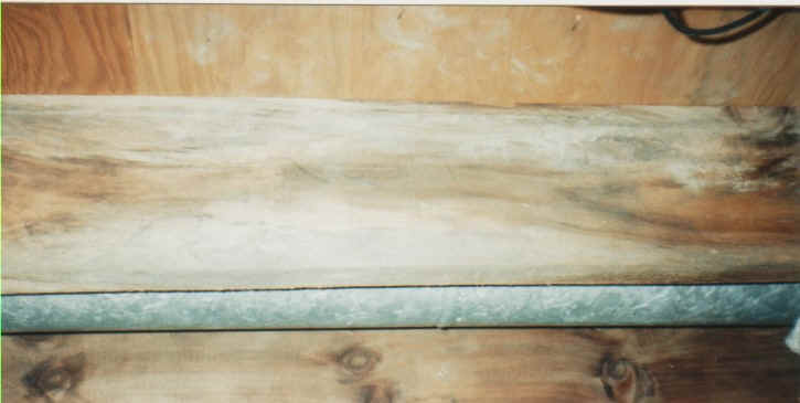
Penicillium & Stachybotrys growing on wallboard in an Oregon apartment
Introduction

Penicillium species are common contaminants on various substances. This organism causes food spoilage, colonizes leather objects and is an indicator organism for dampness indoors. Some species are known to produce mycotoxins. The health of occupants may be adversely affected in an environment that has an amplification of Penicillium.
Species of Penicillium are recognized by their dense brush-like spore-bearing structures. The conidiophores are simple or branched and are terminated by clusters of flask-shaped phialides. The spores (conidia) are produced in dry chains from the tips of the phialides, with the youngest spore at the base of the chain, and are nearly always green. Branching is an important feature for identifying Penicillium species. Some are unbranched and simply bear a cluster of phialides at the top of the stipe. Others may have a cluster of branches, each bearing a cluster of phialides. A third type has branches bearing a second order of branches, bearing in turn a cluster of phialides. These three types of spore bearing systems (penicilli) are called monoverticillate, biverticillate and terverticillate respectively. Penicillium is a large and difficult genus encountered almost everywhere, and usually the most abundant genus of fungi in soils.
The common occurrence of Penicillium species in food is a particular problem. Some species produce toxins and may render food inedible or even dangerous. It is a good practice to discard foods showing the development of any mold. On the other hand some species of Penicillium are beneficial to humans. Cheeses such as Roquefort, Brie, Camembert, Stilton, etc. are ripened with species of Penicillium and are quite safe to eat. The drug penicillin is produced by Penicillium chrysogenum, a commonly occurring mould in most homes.
Penicillium mold growing on the floor joists in the basement ceiling of a home in Massena, New York
Penicillium is characterized by rapidly growing colonies having conidial structures resembling brushes. It commonly produces a strong musty odor. Penicillium marneffei is the only species of the genus that has a yeast-like phase induced by temperature. This can make it more difficult in eradicating it when an infection incurs, but not always impossible.
The antibacterial effect of penicillin was discovered by Alexander Fleming in 1929. He noted that a fungal colony had grown as a contaminant on an agar plate streaked with the bacterium Staphylococcus aureus, and that the bacterial colonies around the fungus were transparent, because their cells were lysing. Fleming had devoted much of his career to finding methods for treating wound infections, and immediately recognised the importance of a fungal metabolite that might be used to control bacteria. The substance was named penicillin, because the fungal contaminant was identified as Penicillium notatum. Fleming found that it was effective against many Gram positive bacteria in laboratory conditions, and he even used locally applied, crude preparations of this substance, from culture filtrates, to control eye infections. However, he could not purify this compound because of its instability, and it was not until the period of the Second World War (1939-1945) that two other British scientists, Florey and Chain, working in the USA, managed to produce the antibiotic on an industrial scale for widespread use. All three scientists shared the Nobel Prize for this work, and rightly so - penicillin rapidly became the "wonder drug" which saved literally millions of lives. It is still a "front line" antibiotic, in common use for some bacterial infections although the development of penicillin-resistance in several pathogenic bacteria now limits the effectiveness.
Growth Media
Commonly found in soil, food, cellulose and grains. It is also found in paint and compost piles. It is also commonly found in carpet, wall paper, and in organic substances inside interior fiberglass duct insulation (NC). Some species can produce mycotoxins. Common cause of extrinsic asthma (immediate-type hypersensitivity: type I). Acute symptoms include edema and bronchi spasms, chronic cases may develop pulmonary emphysema. Many patients complain of a suffocating or gasping sensation when suffering from the effects of penicillium toxicity. But unfortunately, these are also the after effects of many other toxigenic molds.
Penicillium is one of the first fungi to grow on water-damaged materials and has been implicated in causing allergic reactions, hypersensitivity pneumonitis, and a variety of severe lung complications. It may cause sarcoidosis, fibrosis, or allergic alveolitis in susceptible individuals, or patients who have been exposed over long periods of time, depending on the strain.
Toxin Production
Penicillium Aurantiogriseum produces mycotoxins that when ingested or inhaled in large quantities, can cause considerable harm to humans and other mammals. Exposure to large spore loads is to be avoided at all costs. There are a number of toxins reported to be produced by this fungus, and are detailed as follows:
The neurotoxin verrucosidin is produced by this fungus. This toxin was associated with a neurological disease in cattle in the USA (J. Amer. Vet Med. Assoc. 179: 480-81,1991). The mycotoxin penicillic acid is also produced by this organism. Although nephrotoxins, which would cause liver and kidney damage, have been reported to be produced by this mold, the reports may have misidentified the mold. Both cyclopiazonic acid and penicillic acid produced by this mold have acute toxic effects on mammals , and it can be assumed that these mycotoxins are the casual agents of liver and kidney lesions in mice fed with contaminated corn.
Further products include ergosterol and the tremorgenic metabolites tremortin A and B and tremorgen. This mold is further known to produce tropolones puberulic acid and puberulonic acid, a mycotoxin of unknown structure, and a -(L)-malic acid that acts as a proteinase inhibitor. As with all toxigenic fungi, exposure to penicillium does alter human DNA and can cause permanent neurological, pathological, immunological and psychological damage.
Penicillium marneffei produces many serious infections that can be focal or disseminated that can affect the bone marrow, kidneys, lungs, intestines, liver, spleen, skin, and soft tissue. The clinical manifestations most commonly associated with Penicillium marneffei are fever, weight loss, anemia, skin lesions, cough, hepatomegaly, adenopathies, and pulmonary infiltrates.
Apparently, several points of entry are possibly for Penicillium marneffei. The skin, inhalation, and all points of the digestion tract.
Common traits among Penicillium marneffei infected individuals is who have either traveled to and from Southeast Asia and Indonesia, where this fungus is epidemic; and individuals who have been exposed to Penicillium marneffei in water damaged buildings.
This fungal pathogen is unique among Penicillium species as it is the only one to demonstrate a temperature-dependant dimorphic growth stage. A yeast-like dimorphic phase occurs in human tissue and in temperatures at 37C degrees, while at 24C degrees the mycelial phase, which can be quite helpful in devising a plan of action as far as treatments are concerned.
|
Clinical features |
Percentage of cases |
| Fever | 99% |
| Anemia | 78% |
| Weight changes | 76% |
| Skin lesions | 71% |
| Lymphadenopathy | 58% |
| Hepatomegaly | 51% |
| Pulmonary disease/symptom | 49% |
| Diarrhea | 31% |
| Splenomegaly | 16% |
| Oral lesions | 4% |
For treatments, symptoms, associated illnesses, and more information, see
back, for more fungal images and descriptions
Additional Reading
Asan A, Ekmekci S., 1994 The determination of Penicillium and Aspergillus species in Edirne soils and their seasonal distribution Tr J Biol 18:291-303
Ammann, Harriet, Is indoor mold contamination a threat to health?
Bhat RV, Beedu SR, Ramakrishna Y, Munshi KL. Outbreak of trichothecene mycotoxicosis
associated with consumption of mould-damaged wheat products in Kashmir
Valley, India. Lancet. 1989;1:35-37.
[Medline]
Brautbar, Nachman 2002 Toxic molds - The killer within us: Indoor molds and their symptoms
Christensen M, Tuthill DE., 1985 Aspergillus: an overview In: Samson RA, Pitt JI, eds. Advances in Pencillium and Aspergillus systematics. New York: Plenum Press. p 195–209
El-Said AHM., 1994 Studies on soil mycoflora of Bahreen Microbiol Res 149:263-269
Etzel, Ruth, J.A.M.A.; mycotoxins - linking evidence and experience
Filtenborg O, Frisvad JC, Thrane U., 1990 The significance of yeast extract composition on metabolite production in Penicillium In: Samson RA, Pitt JI, eds. Modern concepts in Penicillium and Aspergillus Classification, New York: Plenum Press
Flannigan B, Miller JD., 1994 Health implications of fungi in indoor environments—An overview In: Samson RA, Flannigan B, Flannigan ME, Verhoeff AP, Adan OCG, eds. Health implications of fungi in indoor air environment. 1th ed. Elsevier, Amsterdam.
Forgacs J, Carll WT., 1962 Mycotoxicosis Adv Vet Sci
Fresquez PR., 1990 Fungi associated with soils collected beneath and between pinyon and juniper canopies in New Mexico Great Basin Naturalist 50:167-172
Frisvad JC, Filtenborg O., 1990 Secondary metabolites as consistent criteria in Penicillium taxonomy and a synoptic Penicillium subgenus Penicillium In: Samson RA, Pitt JI, eds. Modern Concepts in Penicillium and Aspergillus Classification. New York: Plenum Press.
Frisvad JC, Thrane U., 1987 Standardised High-Performance Liquid Chromatography of 182 mycotoxins and other fungal metabolites based on alkylphenone retention indices and UV-VIS spectra (Diode Array Detection) J Chrom
Frisvad JC, Thrane U, Filtenborg O., 1998 Role and use of secondary metabolites in fungal taxonomy In: Frisvad JC, Bridge PD, Arora DK, eds. Chemical fungal taxonomy. New York: Marcel Dekker.
Ghildiyal JC., 1993 Mycoflora of decomposing leaf litter in a subtropical freshwater swamp Proc Nat Acad Sci India 63: (B)H 207-211
Gravesen S, Frisvad JC, Samson RA., 1994 Microfungi Copenhagen: Munksgaard
Gravesen S, Nielsen PA, Iversen R, Nielsen KF., 1999 Microfungal contamination of damp buildings—examples of risk constructions and risk materials Env Health Persp 107:Supplement 3.
Gray Michael, 2001 Mold, Mycotoxins and Human Health
Jackson PE, Groopman JD., Aflatoxin and liver cancer. Baillieres Best Pract Res Clin Gastroenterol. 1999;13:545-555. [Medline]
Jesenska Z, Pieckova E, Bernat D., 1992 Heat-resistant fungi in the soil Int J Food Microbiol 16:209-214[Medline]
Khallil AM, Abdel-Sater MA., 1993 Fungi from water, soil, and air polluted by industrial effluents of Manquabad superphosphate factory (Assuit, Egypt) J Basic Microbiol 31:83-100
Khallil A-RMA, El-Hissy FT, Bagy MMK., 1991 Mycoflora of mangroves of Red Sea in Egypt Folia Microbiol 36:456-464
Lillard-Roberts, Susan 2002 Associated illnesses from fungal exposure
Lillard-Roberts, Susan, 2001 Confirming fungal exposure
Lillard-Roberts, Susan 2001, Symptoms of fungal exposure
Marinkovich, Vincent, Sorenson, S.G., Gordon, Wayne A.,Johanning, Eckardt,Haddad, Lisa, Khaboshany, A, Omidi, A, Morsali,S.M., Craner, J.,Stetzenbach, Linda, D., Berek L, Petri IB, Mesterhazy A A, Teren J, Molnar J., Withanage GS, Murata H, Koyama T, Ishiwata I., Pitt JI., Wild CP, Turner PC., Massey TE, Smith GB, Tam AS, Georggiett OC, Muino JC, Montrull H, Brizuela N, Avalos S, Gomez RM., S. Bernardini, G. Falck, A. Hirvonen, H. Järventaus, J. Tuimala, Samson, Robert, A., Kari Reijula, Nolard, nicole, Anna-Liisa Pasanen, Johanning, Eckardt, Landsbergis, Paul, Etzel, Ruth A, Dearborn, Dorr, Ammann, Harriet, Bünger, J., Müller, M., Stalder, K., Hallier E., Medical abstracts on fungal exposure from around the world
Pitt JI.,Toxigenic fungi and mycotoxins. Br Med Bull. 2000;56:184-192. [Medline]
Pitt JI., 1979 The genus Penicillium and its teleomorphic states Eupenicillium and Talaromyces London: Academic Press.
Rayner RW., 1970 A mycological colour chart Kew: Commonwealth Mycological Institute. p 34, charts I and II
Rutherford JM, Huang LH., 1994 A study of fungi of remote sediments in West Virginia caves and a comparison with reported species in the literature NSS Bulletin 56:38-45
Samson RA, Seifert KA., 1985 The ascomycete genus Penicilliopsis and its anamorphs In: Samson RA, Pitt JI, eds. Advances in Penicillium and Aspergillus systematics. New York: Plenum Press.
Smith JE, Solomons G, Lewis C, Anderson JG. Role of mycotoxins in human and animal nutrition and health. Nat Toxins. 1995;3:187-192. [Medline]
Solms-Laubach HG., 1887 Penicilliopsis clavariaeformis, ein neuer javanischer Ascomycet Ann Jard Bot Buitenzorg 6:53-72
Steiman R, Guiraud P, Sage L, Seigle-Murandi F, Lafond J-L., 1995 Mycoflora of soil around the Dead Sea I—Ascomycetes (including Aspergillus and Penicillium), Basidiomycetes, Zygomycetes System Appl Microbiol 18:310-317
Suarez L,
Hendricks KA, Cooper SP, et al. Neural tube defects among Mexican Americans
living on the US-Mexico border. Am J Epidemiol. 2000;152:1017-1023.
Medline]
![]()
©2001-2003 Mold-Help All rights reserved